EKG patterns you may be missing
Andrew Merelman
Twitter: @amerelman
amerelman@gmail.com
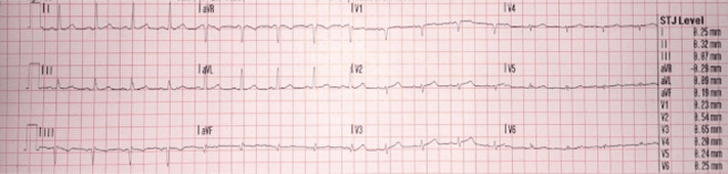
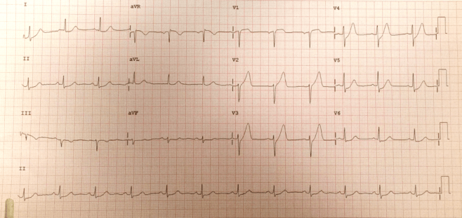
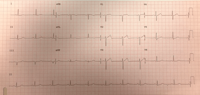
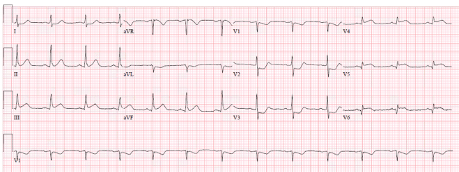
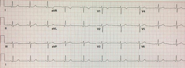
Subtle anterior MI evolving over less than 3 minutes
LAD Occlusion
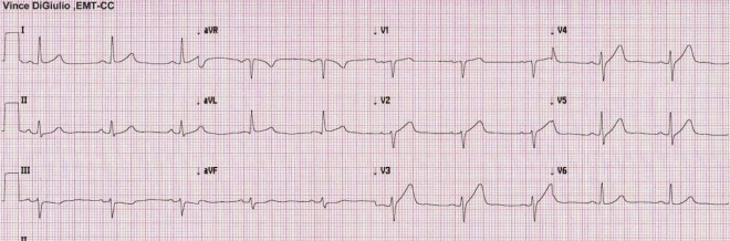
Initial EKG demonstrates subtle anterior MI but was called “normal” by the computer
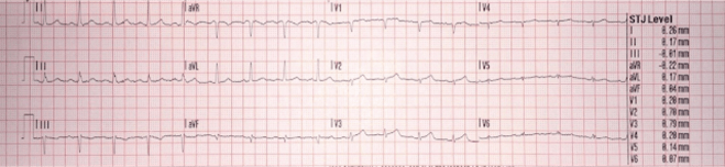
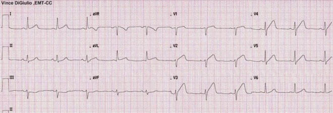
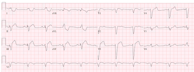
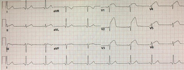
EKG 90 seconds later shows evolution of MI
LAD Occlusion
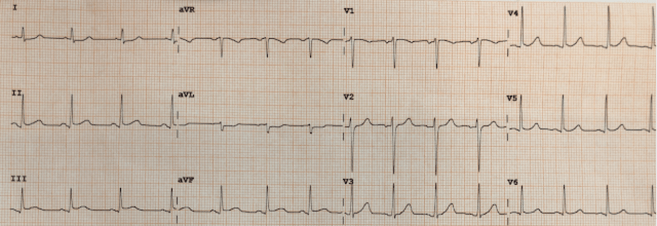
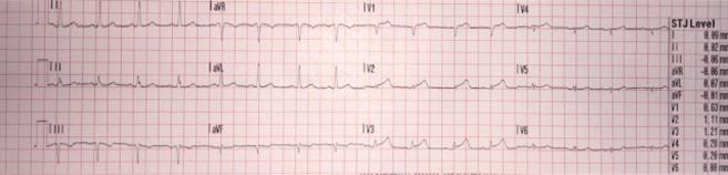
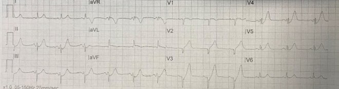
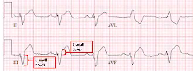
Subtle inferior MI with evolvement
Always look for ST depression and T-wave inversion in aVL, can present BEFORE changes in lead III
RCA Occlusion
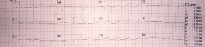
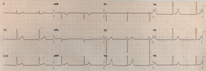
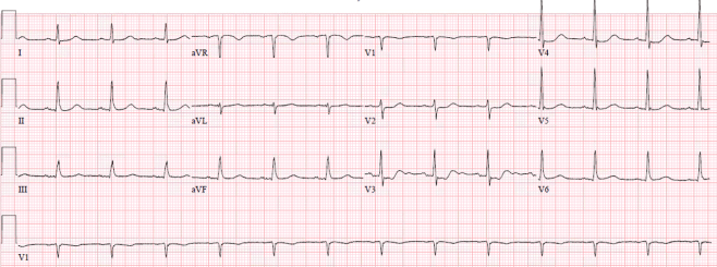
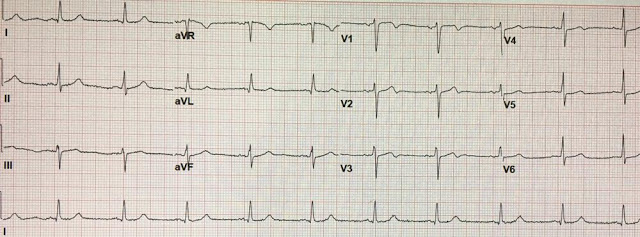
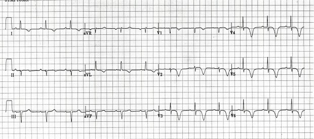
Initial EKG showing de Winter T-waves in leads V2-V5 indicating unstable LAD lesion
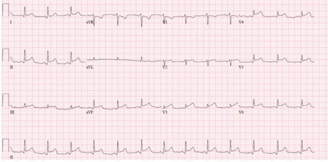
Resolution in second EKG after nitroglycerin, still requires cath lab
Patient had severe LAD stenosis and other lesions requiring CABG
Additional example of DeWinter’s T-waves
Initial EKG shows ST depression in V2-V5 indicating posterior MI
Second EKG shows evolving inferior and lateral MI
The posterior descending artery may arise off the RCA or LCx
This case was due to a LCx occlusion
EKG demonstrating OMI in the setting of LBBB
Use the Modified Sgarbossa Criteria to detect MI in LBBB
Wellen’s Syndrome is when a coronary artery artery occlusion resolves and reperfusion of the vessel occurs. Patients often have chest pain that has resolved at the time of the EKG. Here we see type A Wellen’s where there are biphasic T-waves. The third EKG demonstrates reocclusion of the EKG and was taken during recurrence of chest pain.
https://hqmeded-ecg.blogspot.com/2017/03/subtle-dynamic-t-waves-followed-by-lad.html
Here is an example of type B Wellen’s which would occur similarly to type A. Here we seen deep symmetric T-wave inversions which is typical in reperfusion.
EKG with diffuse ST elevation
This can be mistaken for pericarditis but is actually due to occlusion of a wraparound LAD
Pericarditis is a diagnosis of exclusion, all other life-threatening causes must be ruled out
Most ECGs used (besides de Winter case) are from Dr. Smith’s ECG Blog with permission:
http://hqmeded-ecg.blogspot.com
OMI lecture: https://emcrit.org/emcrit/emcrit-podcast-250-the-omi-manifesto-lecture-by-pendell-meyers/
OMI Manifesto: https://emcrit.org/emcrit/omi-manifesto/
Highly recommend these resources:
“EKG Club” and “12 lead ecg. I’ve got the rhythm” groups on Facebook
http://www.ems12-lead.com
