The myth of the uncontrolled environment

How many times have you heard someone say “you guys work in an uncontrolled environment” as a justification for some deviation from the standard of care? If you work in EMS or the emergency department, i’m guessing quite a few. The ED and prehospital settings are notoriously viewed by others as the Wild West, where we have to do everything as quickly as possible to “save lives”. I’ve seen this mindset throughout my career and it has always bothered me. Why should your “environment” determine the level of care a patient gets? Obviously if a patient cannot be accessed or is physically blocked from an intervention this will dictate care for some period of time. But the vast majority of patients are cared for in open, purposefully designed spaces such as the back of an ambulance or a resuscitation bay. So if the actual space is not the issue, what makes an environment “uncontrolled”?

clint_eastwood_western_1-600x278

I’d suggest that the only thing that makes an environment seem “uncontrolled” is the providers and care team in it. A critically ill patient is the same whether they are in the field, the ED, or the ICU, aside from potentially differing stages of the disease process. The key is how providers prepare for and address issues in their own unique environments. We must be in the mindset that no matter how chaotic or seemingly “uncontrolled” things may be, we cannot degrade the level of care being delivered. Team leaders must be able to ensure the environment remains controlled and things are done properly, no matter the acuity of the patient.

A common example is procedures. Shortcuts are often taken in the emergency setting and there is rarely a reason to do so. We are doing our patients a disservice by knowingly degrading their level of care and putting them at an increased risk of complications. Examples of this include not taking sterile precautions when necessary, not optimizing patients before intubation, and generally rushing procedures due to a perceived time limitation. Many providers have the “faster is better” mentality. But does this actually benefit most patients if the actual procedure is done poorly? I don’t think so.

The corollary is that interventions are often not performed because of the perceived chaos and lack of time. Instead, providers may decide to “let them take care of it later”. This is a common occurrence with prehospital providers delaying treatments and assuming the ED will perform them. And the ED commonly delays treatments assuming the ICU will perform them. These delays can and do impact the quality of patient care.

Recently, I read a comment from a paramedic on an article regarding proper positioning for intubation. The comment suggested it is often impossible to properly position a patient for intubation while in a house, before moving to the ambulance. However, many providers use this as an excuse to not properly position the patient but still perform the intubation. But, the right thing to do, is to not intubate at all until you can properly optimize the patient. As we know, most patients can be managed with basic airway maneuvers until intubation can be performed properly.

Patients should receive the same quality medical care no matter the environment they present. A cardiac arrest patient should receive the same care by paramedics that they would receive if they arrested in the ED, and the patient in the ED should be resuscitated the same as they would be in the ICU. It is our job as emergency and critical care providers to implement processes, perform training, and organize to ensure the sickest patients receive the best care possible.


Please contact me with any questions, concerns, or comments
amerelman@gmail.com
580b57fcd9996e24bc43c53e@amerelman

The myth of the uncontrolled environment

Managing the apneic period – the PreVent trial

Hypoxemia during intubation has long been associated with increased morbidity and mortality. We have had constant struggle to determine the best way to combat peri-intubation hypoxemia. The reality is each patient presents their own oxygenation challenges and may require different approaches. However, one constant is that preoxygenation/denitrogenation is essential for all patients undergoing RSI, though the method may vary.

Picture3

The area of debate has been: how do we handle to apneic period? The primary reason RSI was developed was to prevent aspiration. Therefore, in a customary RSI, no positive-pressure ventilation is provided between induction agent/paralytic administration and laryngoscopy. This is to reduce chances of insufflating the stomach and causing regurgitation. However, many airway experts say their personal practice is to provide gentle BVM ventilation during the apneic period. Some say it is for oxygenation, some say it is more for them to prove the patient can be ventilated should intubation be unsuccessful. These opposing viewpoints have existed without high quality evidence either way, until now.

Untitled

PreVent was a randomized controlled trial performed in seven ICUs in the U.S. They enrolled 401 patients undergoing endotracheal intubation to receive either no positive-pressure ventilation (n=202) or gentle BVM ventilation during their apneic period (n=199). The two groups were remarkably well balanced. The primary outcome was the lowest oxygen saturation from induction to two minutes after intubation.

In the no ventilation group, the median lowest oxygen saturation was 93% vs. 96% in the BVM group. 45 patients (22.8%) in the no ventilation group had severe hypoxemia, defined as oxygen saturation less than 80%, versus 21 patients (10.9%) in the BVM group. These results were all statistically significant. Perhaps more interestingly though, there was no difference in the rate of operator reported aspiration or new opacities on chest x-ray between the two groups.

The authors concluded that BVM ventilation resulted in higher oxygen saturations and lower rates of severe hypoxemia than no ventilation.

Participants received training in BVM ventilation prior to the study. The guidelines were: proper two handed-mask seal, oropharyngeal airway in place, PEEP valve set to 5-10 cmH2O, ventilator rate of 10 bpm, and tidal volumes only large enough to generate chest rise. These parameters demonstrate excellent BVM technique and are an important aspect to the trial.


This was a well-done study with clinically important results. However, there are some important points to consider when determining if this study should change practice:

-The title of the study was interesting to me. BVM ventilation cannot truly be performed during intubation for obvious reasons. This distinction is important because apneic oxygenation can be performed during the apneic period and throughout the intubation procedure.

-Most of the patients in the study were intubated for respiratory failure (~80%). This represents a patient population who are most likely to be affected by our oxygenation decisions during this time period.

-There were significant differences in how the two groups were preoxygenated. The BVM group was much more likely to have BVM used during preoxygenation (40% vs 11%). The no ventilation group was more likely to receive BiPAP (24% vs 16%) or high flow nasal cannula (20% vs 12%). Overall, the BVM group had more preoxygenation with positive pressure (66% vs 55%). It is difficult to say whether the preoxygenation method would have impacted peri-intubation oxygen saturation. Preoxygenation is likely far more important for preventing desaturation than management of the apneic period.

-The no ventilation group received apneic oxygenation 77% of the time, either with an NRB or nasal cannula, although it was not required in either group. If not ventilating the patient, providing apneic oxygenation seems logical, harmless, and potentially beneficial. The authors also do not specify whether or not the airway was maintained during this time period. Apneic oxygenation is not likely to be effective if airway patency is not maintained with a jaw thrust or airway adjunct. Additionally, the BVM group did not receive apneic oxygenation during laryngoscopy, which may have improved outcomes further. Apneic oxygenation has potential to decrease the rate of desaturation during intubation, but the literature is variable.

-Patients who were judged to be a very high risk of desaturation or aspiration, had hypoxemia, or had academia were excluded. These patients are ones that we care most about when it comes to peri-intubation oxygenation and ventilation so it is difficult to say if these results are generalizable to this population.

-The BVM ventilation performed in this study, at least superficially, seems to have been excellent. The guidelines and training were rigid and show that the authors understood the importance of ensuring high quality BVM ventilation to accurately assess the intervention. Unfortunately, this quality of BVM ventilation does not exist commonly in clinical practice. When ventilating during the apneic period, the risk will be greatly increased if poor technique is used.

-The median time from induction agent administration to laryngoscopy in the BVM group was 98 seconds (range 65-135) and 72 seconds (range 52-120) in the no ventilation group. These times are likely longer than typically seen in clinical practice, although most providers are not accurately timing this period so it is difficult to say. The fact that the BVM group had a longer time to laryngoscopy and still had less desaturation supports the conclusion.

-Three of the seven sites used Mapleson F circuits for bag-mask ventilation. These are quite different than a traditional BVM but are likely effective if used by experienced providers. Post from EMCrit on these devices here:
https://emcrit.org/emcrit/what-the-heck-is-a-mapleson-b-circuitu-probably-shouldnt-care/

-Etomidate or propofol were used as the primary induction agent in about 90% of the patients. At induction doses, these drugs are both likely to cause apnea shortly after administration. It is possible that ketamine allows spontaneous respiration for a longer period of time after administration and before onset of paralysis. Since ketamine has become a primary induction agent in the emergency setting, it would be interesting to see if the results would be different had it been used more frequently.

Is this generalizable to the emergency setting?
Since this study was done in ICUs, it is important to determine if the results can be applied to the ED and prehospital setting. One major factor impacting this is whether or not these patients had eaten full meals recently or had been receiving TPN. I could not find anywhere where they address recent food intake in the manuscript. In the emergency setting, our patients almost always have a full stomach. This certainly could impact the rate of aspiration. Another factor is duration of hospitalization. The manuscript does not appear to address how long these patients had been admitted. Patients in the ICU may be in different phases of respiratory illness than typically seen in the emergency setting which could theoretically impact outcomes.

My Conclusions
This is a high quality study which addresses a critical issue in emergency airway management. I think it demonstrates that BVM ventilation is, most importantly, safe during the apneic period and is likely beneficial for preventing desaturation.

How should this be applied to practice?
Patients who are at significant risk of desaturation or are at risk of being harmed be apnea should be ventilated during the apneic period prior to laryngoscopy using EXCELLENT BVM technique with a PEEP valve. This decision should be balanced against the potential risk of aspiration (though it was not demonstrated in this study).

Other Pertinent Clinical Points
-The use of a PEEP valve on the BVM is essential in this setting and is beneficial in almost every other situation as well.

-Apneic oxygenation should be utilized during intubation whether the patient is ventilated prior to laryngoscopy or not. It is a no-risk intervention which has potential to prevent desaturation.

-Preoxygenation is essential prior to any RSI, method may vary.

PLEASE check out Dr. Josh Farkas’ post on PULMCrit discussing this paper and reviewing techniques to manage the apneic period aside from using a BVM. This is essential reading.
https://emcrit.org/pulmcrit/pressure-rsi/

PreVent Trial:
https://www.nejm.org/doi/full/10.1056/NEJMoa1812405?fbclid=IwAR1hpFKkF-t5G2QA2V1Xg_b8MCAXA0LkXPKoxZqoWXYR4LqG58AlQAxwSZM

PreVent Supplemental:
https://www.nejm.org/doi/suppl/10.1056/NEJMoa1812405/suppl_file/nejmoa1812405_appendix.pdf


Please contact me with any questions, concerns, or comments
amerelman@gmail.com
580b57fcd9996e24bc43c53e@amerelman


Casey JD, Janz DR, Russell DW, et al. Bag-Mask Ventilation during Tracheal Intubation of Critically Ill Adults. N Engl J Med. February 2019:NEJMoa1812405.

Managing the apneic period – the PreVent trial

Paralysis without sedation-a negligent act

Imagine this: you are a critically ill patient suffering from sepsis. You are in severe respiratory distress from pneumonia and have become somewhat groggy and slow to answer questions. The medical team treating you appropriately decides that you require intubation to protect your airway and provide ventilatory support. The team properly prepares you for intubation and administers 100 mg ketamine and 70 mg rocuronium. They successfully intubate you and set the mechanical ventilator. At this point, your condition is significantly better as your work of breathing is improved and your airway is protected. But after about 20 minutes on the ventilator you start to regain awareness. You have little recall of the events prior to intubation due to your illness and the use of sedatives. You notice a pain in your throat and want to gag and vomit. You feel that you are breathing but cannot seem to control it yourself. The pain in your throat is worsening to the point where you feel like something is stuck in it. You try to reach toward your mouth but you can’t move at all. Helplessness and pain overcome you and primal fear sets in. You are more aware now than before intubation as the pain has cause you to become alert. The medical team continues to believe that you are comfortable and sedated as you lie motionless on the bed. But they do notice that your heart rate has increased substantially from 120 prior to intubation to 180 now. They believe this is due to your illness and the negative effects of positive pressure ventilation, so they administer more IV fluid. Only after an hour do you regain the ability to breathe or move. You start to over-breathe the ventilator and set off alarms. You reach for your endotracheal tube and luckily a nurse stops you from pulling it. Only now are you given sedation and fall back into unawareness.

IMG_1915

Sedation and analgesia of intubated patients in the emergency department is historically poor. In my experience, this is due to several factors:

  1. A misunderstanding of paralytic medications
    Neuromuscular blockers do not provide any sedation or pain relief.
    I’m going to repeat that: neuromuscular blockers DO NOT provide any sedation-or analgesia.
    I could give you 70 mg of rocuronium, 7 mg of vecuronium, or 150 mg of succinylcholine right now and, after about 30-60 seconds, you would become unable to move, breathe, or even blink, but would remain completely conscious. This means that any patient receiving a long or short acting paralytic MUST receive sedation as well, at least for as long as the duration of that paralytic.
  2. A misunderstanding of the duration of induction agents
    After intubation in the emergency department, it is typical for providers to let their guard down, believing the hard work is done. This often looks like people leaving the room or becoming focused on other tasks. This is a huge problem for many reasons, as patients are at there most vulnerable during AND immediately after intubation. One of the problems with this is that post-intubation analgesia and sedation is not made a priority. The most common induction agent used in the ED is etomidate. This drug provides adequate sedation for maybe 3-5 minutes. Unless you have prepared post-intubation sedation drugs prior to intubation, it is likely there will be a gap in sedation. If the patient has received a long-acting neuromuscular blocker (most commonly rocuronium), this means the patient may have a period of awareness, in which paralysis continues despite a return to consciousness. Even succinylcholine, which has a shorter duration, can still last up to 10 minutes, depending on the patient. So even then, the patient could have a period of awareness.
    The best way to avoid this is to ensure post-intubation sedation is prepared PRIOR to intubation in EVERY patient, regardless of induction agent.
  3. A fear of sedating critically ill or hemodynamically tenuous patients
    The sedatives frequently used in the emergency setting are known to cause decreases in blood pressure (e.g propofol, midazolam, lorazepam). In very ill patients, this can cause problems. However, there is almost never a time where a patient is so hemodynamically compromised that they cannot receive any analgesia or sedation. This is especially true with modern post-intubation analgosedation practices. This includes the idea of providing analgesia first, typically fentanyl, which can reduce the amount of sedative required and likely result in improve hemodynamics over sedatives alone. Additionally, ketamine is an effective post-intubation agent and is more likely to maintain hemodynamic parameters than other agents. It has the added benefit of providing analgesia in addition to sedation (dissociation). Also, It is acceptable to initiate or increase vasopressors if necessary to support blood pressure. Hemodynamic compromise is not an adequate excuse for poor sedation.
  4. Laziness
    This is a problem that may be more prevalent in the prehospital setting simply due to the regulation of controlled substances and more strict documentation requirements, but can certainly be a factor in the emergency department as well. Using controlled substances often requires additional paperwork, time, and effort which a busy emergency provider may not want to deal with. This is clearly unacceptable but is a real occurrence.

Post-intubation best practices:
-Prepare all necessary agents prior to intubation
-Use an analgesia first strategy, typically with fentanyl or ketamine (at analgesic or dissociative doses). Hydromorphone can also be used for this purpose.
-Monitor for signs of awareness – increased HR, BP, or ETCO2 can be seen even in paralyzed patients
-Consider ketamine as a sole agent for post-intubation sedation in the emergency setting in infusion and/or bolus form. It provides rapid, reliable sedation (dissociation) with a longer duration of action than traditional agents. It is also more hemodynamically neutral than other drugs (and potentially sympathomimetic) making it the agent of choice in tenuous patients.
-Use vasopressors as necessary to support post-intubation hemodynamics and to facilitate adequate analgosedation.

For a modern approach to post-intubation sedation and analgesia see this article:
https://www.emsworld.com/article/220258/a1-sedation-package-better-care-intubated-patients

Co-authored by Michael Perlmutter
580b57fcd9996e24bc43c53e@DitchDoc14

Please contact me with any questions, concerns, or comments
amerelman@gmail.com
580b57fcd9996e24bc43c53e@amerelman

References


  1. Long-acting neuromuscular paralysis without concurrent sedation in emergency care. Chong ID, Sandefur BJ, Rimmelin DE, Arbelaez C, Brown CA 3rd, Walls RM, Pallin DJ. Am J Emerg Med. 2014 May;32(5):452-6. doi: 10.1016/j.ajem.2014.01.002. Epub 2014 Jan 15.

  2. Estimates of sedation in patients undergoing endotracheal intubation in US EDs. Weingart GS, Carlson JN, Callaway CW, Frank R, Wang HE. Am J Emerg Med. 2013 Jan;31(1):222-6. doi: 10.1016/j.ajem.2012.05.015. Epub 2012 Jul 4.

  3. Prevention of awakening signs after rapid-sequence intubation: a randomized study. Jaffrelot M, Jendrin J, Floch Y, Lockey D, Jabre P, Vergne M, Lapostolle F, Galinski M, Adnet F. Am J Emerg Med. 2007 Jun;25(5):529-34.

  4. Inadequate provision of postintubation anxiolysis and analgesia in the ED. Bonomo JB, Butler AS, Lindsell CJ, Venkat A. Am J Emerg Med. 2008 May;26(4):469-72. doi: 10.1016/j.ajem.2007.05.024.

  5. Rapid Sequence Intubation from the Patient’s Perspective. Kimball D, Kincaide RC, Ives C, Henderson S. West J Emerg Med. 2011 Nov;12(4):365-7. doi: 10.5811/westjem.2010.11.1922.

  6. 5th National Audit Project (NAP5) on accidental awareness during general anaesthesia: summary of main findings and risk factors. Pandit JJ, Andrade J, Bogod DG, Hitchman JM, Jonker WR, Lucas N, Mackay JH, Nimmo AF, O’Connor K, O’Sullivan EP, Paul RG, Palmer JH, Plaat F, Radcliffe JJ, Sury MR, Torevell HE, Wang M, Hainsworth J, Cook TM; Royal College of Anaesthetists; Association of Anaesthetists of Great Britain and Ireland.

  7. Weingart S. EMCrit Podcast 21—A Bad Sedation Package Leaves Your Patient Trapped in a Nightmare. EMCrit RACC, https://emcrit.org/racc/post-intubation-sedation/.
  8. Scott Weingart. Podcast 115 – A New Paradigm for Post-Intubation Pain, Agitation, and Delirium (PAD). EMCrit Blog. Published on January 13, 2014.
  9. Early deep sedation is associated with decreased in-hospital and two-year follow-up survival. Balzer F, Weiß B, Kumpf O, Treskatsch S, Spies C, Wernecke KD, Krannich A, Kastrup M. Crit Care. 2015 Apr 28;19:197. doi: 10.1186/s13054-015-0929-2.

  10. Comparative evaluation of intravenous agents for rapid sequence induction–thiopental, ketamine, and midazolam. White PF. Anesthesiology. 1982 Oct;57(4):279-84.

  11. Comfort and patient-centred care without excessive sedation: the eCASH concept. Vincent JL, Shehabi Y, Walsh TS, Pandharipande PP, Ball JA, Spronk P, Longrois D, Strøm T, Conti G, Funk GC, Badenes R, Mantz J, Spies C, Takala J. Intensive Care Med. 2016 Jun;42(6):962-71. doi: 10.1007/s00134-016-4297-4. Epub 2016 Apr 13. Review.


Paralysis without sedation-a negligent act

Why we need more go-arounds in medicine

While landing an aircraft, a pilot may decide that the landing has become unsafe or unlikely to be successful. This may be due to a poor approach, another aircraft on the runway, deteriorating weather, or numerous other reasons. Standard procedure in this case is to abort the landing attempt, which involves advancing the throttle to full power, retracting the flaps and landing gear, and establishing a positive rate of climb. The pilot announces to the control tower that they are “going around”. The control tower calmly responds with instructions for the aircraft to set up its repeat approach.
500px-Go-around
This procedure is well established and commonplace in aviation. As all pilots have, I practiced this procedure many times during my initial flight training and still practice it when I fly. A good pilot is compelled to go around whenever there is any question about the ability to land safely and because of this, a pilot’s decision to do so is never questioned. However, a go-around itself can be dangerous and must be performed properly, as the aircraft is going from low speed into a climb, which increases the risk for stall.

Here is a great video of a go-around. You will hear the ground-position warning system announce “approaching decision height” and then “minimums”. The decision height is the altitude the pilot must decide whether to continue the approach. Minimums indicates that the aircraft is at the altitude where the pilot must be able to visualize the runway to continue the approach.

The pilots had a usable view of the runway and initially continued their approach. Immediately after reaching minimums, the rain became so heavy they could no longer see the runway to continue the landing. They had to make a rapid decision to go around and completely change their mindset from landing to establishing a climb with no visibility, on instruments, and with autopilot off. This is a massive cognitive burden. But there is no panic, argument, or discussion. There is only procedure. These pilots have practiced this before. They know it is the right decision and that no one will criticize it.

In medicine, we have a huge problem admitting when things are not going as planned, especially if we are the ones who made that plan. We worry that we will be scrutinized by others or that we will appear weak.
Imagine a scenario where you are performing a rapid sequence intubation. You are the one intubating and the provider directing the patient care team. You made sure the patient was well prepared, preoxygenated, and hemodynamically supported. You’ve established an oxygen desaturation threshold of 93% to discontinue the attempt. The drugs go in, the patient is paralyzed. You insert the blade as you always do but you don’t see what you are expecting. The epiglottis is visible but it won’t lift. This is the point where your heart rate increases. The moment things begin to deviate from the expected course is the moment situations deteriorate. You try to readjust your blade but the view only worsens. You try to use your Mac as a Miller but can’t see anything after progressing past the epiglottis. You back the blade up again and finally think you can see a sliver of the posterior cartilages, but its not enough. You think you just need to make one final adjustment to have a usable view and someone says, “the sats are 93”.
This is “minimums” in airway management. Do you make that final adjustment to see if it works? Or do you abandon the attempt and ventilate the patient? As with aviation, this decision must be made with safety in mind.
So why is it that so often we reach “minimums” but make the wrong decision? A decision made, not in the name of safety, but out of pride or fear of scrutiny. Or is it because we truly think it is the right thing to do for the patient? This decision is rooted in a culture of task fixation. This culture is instilled in us by our training from day one. How often do we practice abandoning an intubation attempt and reoxygenating a patient? This practice is less about the actual skill and more about how and when to make the go-around decision.

The idea of a go-around is not strictly applicable to airway management. This applies anytime patient care is not going as planned or the patient has a sudden deterioration and requires a rapid change in treatment.

We must:
1. Know and teach how to recognize deteriorating situations or procedures that are unlikely to be successful
2. Know and teach how to respond to these situations, our go-around procedure
3. PRACTICE our go-around procedures as regularly as we practice other treatments
4. Be prepared to change our approach at the drop of a hat if patient safety is at stake

This is about changing culture. We must encourage more go-arounds during resuscitation. Because in many cases, if we don’t go around, patients die.

Here’s what happens when you don’t go around in aviation when you should.
1200px-Nose_and_forward_section_of_UPS_1354
NTSB: “The flight crew did not monitor the descent rate and continued to fly the airplane with a vertical descent rate of 1,500 ft per minute below 1,000 ft above ground level, which was contrary to standard operating procedures, resulting in an unstabilized approach that should have necessitated a go-around.”

Reviewed and edited by Michael Perlmutter
580b57fcd9996e24bc43c53e@DitchDoc14

Please contact me with any questions, concerns, or comments
amerelman@gmail.com
580b57fcd9996e24bc43c53e@amerelman

Why we need more go-arounds in medicine

Why I use a bougie on every airway

The bougie, often referred to as the gum-elastic bougie, is neither gum-elastic nor a bougie. It is probably best to call it an endotracheal tube introducer but it is widely known as the bougie. Nonetheless it is a device essential to emergency airway management.

During my initial paramedic training, I received very little education on the bougie. My love for this device has only come through my own realizations as I have expanded my airway knowledge through FOAMed and advanced airway courses. Also, my good friend Jorge Cabrera (@In10sivist), is a bougie fanatic and has taught me a great deal about the nuances of this tool. The traditional view of the bougie is that it is a “rescue” device and is only needed in challenging airway situations. In fact, many providers view the bougie as a form of “cheating” and imply that if you use it you have, in some way, failed. This mindset is not only ridiculous, it is dangerous, and discourages the application of a safe device that can really save you and your patient.bougieSo why is a bougie so useful? The objective answer is that it allows you to intubate patients with a more limited glottic view. As a general rule, it is best not to try to pass a styletted tube with a view that is worse than grade 2A. This is because anything worse does not allow you to visualize the tube entering the cords. Additionally, an ET tube is large and often obscures your view of the cords while trying to maneuver it to them.

5266270103_6cc150f80b
Nurse-anesthesia.com

A grade 2A view is the minimum safe view that should be used for a styletted tube. But with a bougie, you can now safely and reliably deliver a tube with a grade 3A view. The coudé tip can be carefully passed beneath the epiglottis and up into 3avs3bthe larynx. This is obviously not optimal, as it is always best to try to visualize the cords. But if you can only see epiglottis after performing epiglottoscopy, elevating the head, doing external laryngeal manipulation, and using your mac blade as a miller, you do not have to abandon your attempt.

You may be asking, “but couldn’t you just have a bougie ready and only use it if you need it?” Of course the answer is yes, but why not use the device most likely to be successful on every intubation? The modern airway approach is designed to achieve first-pass success. If you plan to use an ET tube with a stylette it is probable that you will try this approach first, regardless of whether your view is adequate. You are psychologically more likely to attempt your initial plan even if you should not. This means that you may end up wasting time jabbing a tube around the pharynx, causing trauma, and prolonging your attempt.
It is also markedly easier to pass a bougie through the cords and down the trachea than it is to pass a tube. Even with a perfect view of the larynx you can still run into issues advancing the tube. The primary reason for this is impacting the cricoid ring with the tube tip. This can often be overcome by rotating the tube 90° clockwise, but why risk it? Passing a bougie gives you a guide into the trachea. It is the Seldinger technique of

rotate-Rural Doctors Net
Rural Doctors Net

airway management. Once it is placed you have a reliable route to deliver your ET tube. “But what if the tube gets caught on the cords?” This is a relatively common, but easily overcome issue. If the tube is passed with the bevel left, as most tubes are designed to be by default, the tip may get caught on the right vocal fold. This is resolved by simply rotating the tube 90° counter-clockwise. This puts the bevel down and allows the tip to pass between the cords. My preference is to rotate the tube as I advance it so this is never an issue.

But there are other advantages to the bougie. The best part for me is the instant placement confirmation. You have immediate feedback about whether the bougie is in the trachea or the esophagus in the form of tracheal clicks. As the coudé tip advances down the trachea, it rubs against the tracheal rings. This is palpable as clicks felt when gripping the bougie. Many will say that these cannot always be felt.

1-s2.0-S0735675704001871-gr3
Phelan, 2004

ANECDOTE AHEAD: In forty two cadavers and many live intubations I have been able to feel tracheal clicks in every person. This is not to say there aren’t patients that clicks cannot be felt in. The key is ensuring the coudé tip remains pointing upward. If the tip rotates to the side or downward, it will not be against tracheal rings and the clicks will not be felt. You also must advance the bougie slowly (for many reasons) and consciously feel for clicks, as sometimes they are subtle. Additionally, you can ask an assistant to place their hand on the trachea and they will also be able to feel the clicks.
The other way that placement can be confirmed is by achieving hold-up in one of the proximal bronchi. If the bougie has been placed properly, it will only advance a short distance before impacting the carina and then lodging in the bronchi. If it is placed in the esophagus, it will not stop advancing. This method is somewhat controversial due to the risk of tracheobronchial trauma. This risk can be mitigated by advancing the bougie very slowly when attempting to achieve hold-up. However, my personal approach is not to achieve hold-up if I have felt tracheal clicks. I will advance far enough that the bougie will not dislodge from the trachea and stop. This avoids the risk of airway trauma. But, if clicks are not felt and you believe the bougie is in the right place, advancing slowly and achieving hold-up is a reliable way to confirm placement.

The last reason I use a bougie on every intubation is familiarity. The more you perform a skill the better you will be at it, especially in live patients. Using the bougie on “easy” intubations increases familiarity and experience with the device. This way, when you really need it, you will be comfortable handling it and know what to expect.
I view the bougie as the stylette is viewed now. When stylettes first became mainstream, there was stigma against them and there was a view that using them was “cheating”. Today, in most modern countries, the stylette has become the standard of care and intubating without it is seen as inferior because of its ability to make tube delivery significantly easier.

Video: Sam Ghali, @EM_Resus

Bougie Tips
A common error is removing the laryngoscope from the mouth once the bougie is placed. Your view of the larynx, however excellent or restricted it is, should be maintained until the tube is placed. There are two reasons for this. Number one is to ensure the bougie does not dislodge and to try to visualize the tube entering the trachea to confirm it goes in the right place. The other is that removing the laryngoscope causes the upper airway to collapse on the bougie making it more difficult to advance the tube. This can also cause bougie dislodgement if it is not advanced far enough into the trachea.

The bougie is generally not recommended when using indirect video laryngoscopes (i.e standard Glidescope, CMAC D-Blade). These are devices that can only be used for video laryngoscopy and are not shaped to perform direct laryngoscopy. The reason for this is that they do not displace airway tissue and create a steep angle to the cords. The best way to deliver tubes when using these devices is a hyperangulated stylette with a 60-70° bend that matches the bend of the blade.

shaka
“Shaka” grip (airwaycam)

There are many techniques out there involving preloading the ET tube and different grips. I prefer to use the bougie “naked” without preloading the tube. I feel that it gives me more control and better ability to feel trachea clicks. The tube is then advanced by another provider over the bougie or I hold the bougie against the laryngoscope with my left hand and load the tube myself with my right hand. The tube is then advanced and passed between the cords. This is not evidence based and many techniques may be equally effective. Practice all of them and find one that works best for you. The Shaka grip gives you the ability to elevate the tip of the bougie with your pinky which may help maneuver below the epiglottis. It also provides a direct indication of which way the coudé tip is pointing to ensure it stays upright. There are many different ways to preload a tube including the D-grip or Kiwi grip.

photo-32
Preloaded bougie (Prehospitalmed.com)
Kiwi grip airwaycam.com
“Kiwi” grip (airwaycam)

The bougie can be especially useful with an Omega shaped epiglottis. It can be difficult to pass a tube but the epiglottis acts as a conduit for the bougie.
(Videos: Sam Ghali, @EM_Resus)

Quick points because of things I’ve seen attempted:
-Do not use straight (non-coudé) tip bougies. They will not generate tracheal clicks and may be more likely to cause trauma as they will advance further into the tracheobronchial tree.
-Do not attempt to blindly place a bougie. It is not a heat-seeking missile.
-The smallest tube that will fit over an adult bougie is 6.0 mm, pediatric 3.5 mm.

Feel free to contact me with any comments, questions, or concerns
amerelman@gmail.com
580b57fcd9996e24bc43c53e@amerelman

References

Airway FOAMed
http://www.emdocs.net/novel-tips-airway-management/
https://lifeinthefastlane.com/ccc/bougie/

Driver BE, Prekker ME, Klein LR, Reardon RF, Miner JR, Fagerstrom ET, Cleghorn MR, McGill JW, Cole JB. JAMA. 2018 Jun 5;319(21):2179-2189. doi: 10.1001/jama.2018.6496.

Driver, B., et al. (2017). The Bougie and First-Pass Success in the Emergency Department. Annals of Emergency Medicine. doi:http://dx.doi.org/10.1016/j.annemergmed.2017.04.033

Kidd, J. (1988). Successful difficult intubation. Use of the gum elastic bougie. Anaesthesia, 43(6).
doi:10.1111/j.1365-2044.1988.tb06625.x

Gataure, P., et al. (1996). Simulated difficult intubation. Comparison of the gum elastic bougie and the stylet. Anaesthesia, 51(10).
doi:10.1111/j.1365-2044.1996.tb14961.x

Phelan, M. (2004). Use of the endotracheal bougie introducer for difficult intubations. The American Journal of Emergency Medicine, 22(6). doi:https://doi.org/10.1016/j.ajem.2004.07.017

Why I use a bougie on every airway

Taming the bag-valve-mask

Using a BVM *properly* is, without a doubt, one of the most challenging tasks we perform in EM, EMS, and critical care. Most providers do not get enough initial training or ongoing practice. Perhaps the biggest factor that makes people do this poorly is the sympathetic surge experienced while ventilating a patient. This leads to lack of focus on the task and poor quality ventilation. It requires calm and collected performance when the brain is anything but. The person ventilating must be absolutely focused on that task and not distracted by other issues. Its not all our fault though. The BVM is really nothing more than a bellows reshaped to fit on people’s face, not the most advanced device.


-The first step to good BVM technique is properly positioning the patient. See my last post here for information on that topic.-

1. The Mask Seal
A good mask seal is essential for allowing the BVM to work at its full potential. Leaks lead to inadequate ventilation and loss of airway pressure between breaths. There are a few ways to maintain an adequate seal. Please note: the mask seal should be maintained at all times and not interrupted in between breaths. This allows the maintenance of airway pressure even during exhalation and between breaths. It is important to maintain airway pressure. Oxygenation is maximized with increased mean airway pressure.
When performing one-person BVM you can use the C-E grip to maintain a jaw thrust and mask seal. The non-dominant hand should be used to maintain a seal. The thumb sits on the nose side of the mask and the index finger wraps around the bottom of the mask. The other three fingers are placed on the jaw bone with the pinky at the back of the jaw. These fingers should pull the jaw forward maintaining a jaw thrust. The fingers on the mask should be used to help maintain the seal and minimize leaks. It may help to use the bag portion of the BVM as a lever to provide more mask seal on the side of the mask that is not being held. The bag can be pushed downward resulting in the mask being pressed into the face more on that side.
IMG_9160The optimal way to perform BVM ventilation is with two providers. A mask seal is held with both hands by one provider and the other squeezes the bag. When maintaining a mask seal with two hands a double C-E grip can be used. This allows both hands to be used for displacing the jaw forward and results in significantly improved mask seal. Another, often more effective, technique is placing the palms of both hands on the sides of the mask then using the index and other fingers to pull the jaw forward. This method may be preferred in difficult BVM situations. Maintaining a jaw thrust is essential to maximizing oxygenation. Otherwise the airway obstructs and prevents air passage.

2. Squeezing the bag
This part is important and can really make your patients worse if it is done poorly. The typical adult BVM has a volume of 1.0-1.2 liters. The tidal volume desired is usually about half of that. This means that you DO NOT need two hands to squeeze the bag. One hand is plenty sufficient and, in most cases, you can use two fingers. Only enough volume to cause chest rise and ETCO2 return is needed. Some people say to even use a pediatric BVM for adults because it is much closer to the actual tidal volume necessary. If the patient is spontaneously breathing simply augment the patient’s own breaths with a small volume. So why is volume so important? Too much volume can lead to barotrauma so it is important to avoid this. Also, providing too much volume results in hyperinflation of the lungs, increased intrathoracic pressure, and decreased venous blood return to the heart. Most sick patients rely on adequate preload so killing it with the BVM can really hurt them.
Volume is only part of the story though. We also have to be cognizant of the amount of pressure we deliver, the speed of the squeeze. There are a few reasons for this. When delivering breaths with a mask, as opposed to an ETT tube or SGA, air can go two places. The place it likes to go most is the lungs as there is not much resistance in that pathway. However, the lower esophageal sphincter can be overridden with only  a small amount of pressure. This results in gastric distention. It only takes a short time to completely fill the stomach with air and distend it significantly. This hurts us, and the patient, in multiple ways. The first is that people tend to vomit when their stomach is filled with air. This make airway management and ventilation more challenging. Additionally, filling the stomach with air causes it to compress the diaphragm and inhibit lung expansion which further impedes ventilation. So how can you minimize this? Make sure you deliver breaths slowly, over at least two seconds, if not longer. You can also use a pop-off valve that limits the amount of pressure that can be delivered.
The last part of the story is the rate. Now this is where people get really excited and make their patients sicker. There are very few patients that need 40 breaths/minute. It is important to consciously maintain an appropriate ventilatory rate. This is easily done by monitoring ETCO2. Shoot for a number that is appropriate for the patient condition, normal is 35-45 mmHg. If you’re going to fast it will decrease, too slow and it will increase.
In summary, deliver small volumes, with low pressures, at slower rates and this will ultimately benefit your patient.

3. Use airway adjuncts
Basic airway adjuncts can go a long way in the difficult to ventilate patient. In reality though, if you use all the tips in this post, you usually will not need any basic adjuncts. However, some people have large tongues and extra soft tissue that cannot be displaced with simple positioning and jaw thrust. In completely obtunded or unresponsive patients it is prudent to insert an adjunct initially to maximize chances of successful ventilation. Inserting a properly sized nasopharyngeal airway or oropharyngeal airway helps to bypass the tongue and create a passage for ventilation. Keep in mind the device must be properly sized so that it reached past the base of the tongue. If it does not reach far enough then all it is doing is acting as an obstruction and making ventilation more difficult. Also, keep in mind that inserting either device can illicit the gag reflex leading to vomiting.

B9781455705542000058_f005-011-9781455705542
Image: pocketdentistry.com

4. PEEP
All aspects of airway management and assisted ventilation involve PEEP. Historically, PEEP use with a BVM has been minimal but recently it has become standard of care. PEEP (positive end expiratory pressure) is the amount of pressure that is maintained in the lungs and airways at the end of exhalation. This pressure is maintained by the glottis and upper airway structures in normal physiology. This pressure is what allows the alveoli to remain inflated and not collapse during the exhalation phase. When alveoli collapse, also known as atelectasis, there are a few adverse IMG_91601effects. The first is that they become significantly harder to recruit and inflate. Once an alveoli is collapsed it requires much more pressure to reinflate it. Alveoli that are collapsed cannot perform gas exchange leading to worsened oxygenation and ventilation. Additionally, when atelectasis occurs alveoli become damaged, less effective, and may rupture. This is especially true in patients with lung disease. Because of this, a PEEP valve should be used on all BVMs and adjusted individually for each patient. The typical setting for healthy lungs is 5 CMH2O but this can be increased in certain situations. Patients with pulmonary edema or other causes of physiologic shunt often require more PEEP to oxygenate and recruit lung tissue. Do not be afraid to increase PEEP if the oxygen saturation is not improving and always use at least 5 CMH2O.
PEEP can also aid in ventilation. Maintaining higher airway pressures, in combination with jaw thrust and good technique, can help keep the airway patent and maximize air movement. In order for PEEP to be effective the mask seal must be maintained at all times, even in between breaths.
Below are two videos from George Kovacs (@kovacsgj) that he developed in one of his cadaver labs. They demonstrate the incredible effects of PEEP and why it is so important.

BVM with PEEP

BVM with ETT and PEEP

5. BVM as CPAP/BiPAP
The application of PEEP via a BVM has another advantage. In the spontaneously breathing patient the BVM can be used as CPAP or BiPAP. If the mask is sealed well on the face, at least 15 lpm oxygen is flowing, and a PEEP valve is in place, the patient will receive the set amount of PEEP in the form of CPAP. Additionally, if you squeeze the bag when the patient breaths you can essentially provide BiPAP. Spontaneously breathing patients, even if minimally, often benefit greatly from only CPAP via BVM without squeezing the bag. This decreases the risk of gastric insufflation while providing support to the patient’s own respiratory drive.
Also, placing a nasal cannula under the mask at 15 lpm to provide additional oxygenation. It also generates additional airway pressure which supports the generation of PEEP. This is an excellent technique to use for preoxygenation prior to intubation without having to setup a CPAP or BiPAP machine.
Video below, also from George Kovacs, demonstrates this technique.

PEEP as CPAP

6. Add a nasal cannula
The nasal cannula has become a mainstay of airway management. Oxygenation through the nose is significantly easier and more effective than through the mouth. Adding a nasal cannula at 15 lpm or greater under the BVM has great benefit. It increases the overall FiO2 delivered and it aids in generating airway pressure when combined with a PEEP valve.
You can also give apneic CPAP during the apneic period of RSI. Patients who require PEEP to oxygenate should have it maintained for as long as possible without interruption. Once the airway pressure decreases the alveolar recruitment generated by the PEEP is lost. But, during RSI, we often try to avoid ventilating during the apneic period for fear of regurgitation. PEEP is usually generated by breathing or ventilating but is typically lost during apnea. However, adding the nasal cannula allows PEEP to be maintained as it provides flow inward which increases airway pressure. With this, you can maintain your BVM mask seal during the apneic period and help maintain airway pressure without ventilating.

7. End-tidal CO2
ETCO2 should be used on all patients who are obtunded or have respiratory distress. It is an invaluable tool for monitoring respiratory status. It can be done with a nasal cannula type device or in-line device. An in-line ETCO2 adapter can be placed between the mask and the BVM adapter in the same way it would be placed on an ETT. Maintain a good mask seal and you will get a nice ETCO2 waveform to help guide your ventilation. If you are not getting a waveform this is indicative of poor mask seal or lack of air movement through the airway. If this occurs adjust mask seal and ensure the jaw is being pulled forward.
normalcapnograph
Summary
The BVM is a difficult device to master. Whenever you use it be sure to consciously consider HOW you are using it. Position the patient properly, upright and ear-to-sternal notch. Always make sure to maintain a constant mask seal. Deliver small, low pressure breaths. Use airway adjuncts as needed. Add a PEEP valve to maximize alveolar function and consider using the BVM for CPAP or BiPAP. Add a nasal cannula with 15 lpm O2. And finally, always use ETCO2 when ventilating a patient.

Remember: if this guy can do it, so can you.

Like us on Facebook!

Questions, comments, feedback?
theresusroom@gmail.com
@amerelman on twitter

Taming the bag-valve-mask

Airway anatomy – and how we make it worse

“The best position to kill a patient in, from an airway perspective, is flat on their back.”
-Rich Levitan – @airwaycam

This is one of the most important concepts in emergency airway management. Rich knowszTXey5ETB this better than anyone, having examined 20 cadaveric airways per month for the past 16 years in addition to clinical research. He coined the term “coffin position” to describe the detriment of having your patient supine. Cannot Oxygenate Face Flat in Neutral position. There are a few reasons this is true and it is important to understand them. Not only will it help improve patient outcomes but it will make you better at managing airways.

The Upper Airway
When we talk about airway management we are talking about the upper airway. Barring any severe trauma or pathophysiology, the airway south of the glottic opening is intrinsically patent. We are really only worried about the passageway above the glottis which is made up of soft tissues that do not have spontaneous patency. Normally, patency is maintained by muscles controlled consciously and unconsciously. But when patients are obtunded or sedated they lose the ability to maintain upper airway patency.

Untitled
Graphic: airwaycam.com
People do not maintain a perfectly straight posture. The neck curves forward which maintains the head in a slightly forward position. From an airway perspective, this helps keep the tongue and other soft tissue off of the posterior pharynx. You can easily see how this is effected when you stand flat against a wall. You will see that placing your head straight back against the wall causes discomfort. This shortens the thyromental distance leaving less room for the tongue and decrease mouth opening. Obviously you can still breath as you are able to maintain conscious control of your airway. But compromised patients lose this ability when they are positioned this way. Now, take the a patient and lay them flat on their back. Not only do you misalign the upper airway but the tongue and soft tissues fall back with gravity. These two mechanisms will undoubtedly cause partial or complete airway obstruction in obtunded patients.

Airway xray
Left: Ear-to-sternal notch, Middle: Neutral, Right: Hyperextended                    Graphic: Rich Levitan
The Lungs
Bad things happen to the lungs when a patient as lying flat. It becomes much more difficult to fully recruit lung tissue. This is due to a number of factors and is much more profound in obese patients. The abdominal contents are no longer held off the diaphragm by gravity. They move upward and often push on the diaphragm preventing it from contracting fully. This also decreases the amount of thoracic space the lungs have to expand in. The net result is decreased amounts of usable lung tissue, lower functional residual capacity, and more difficulty recruiting alveoli. In addition, the obese have weight from fat which puts pressure on the chest and abdomen preventing lung expansion and contributing to the abdominal component of supine positioning. Obese patients desaturate significantly faster than average sized patients, mainly for this reason.

The Stomach
RSI is a technique that was designed with the stomach in mind, not the lungs. This is very important as it highlights one of the most detrimental complications during airway management, regurgitation. Vomiting can be active or passive. In an RSI scenario it is often passive as paralytics are given to help prevent it. However, active or passive vomiting can occur at any time during the airway management process. It is further encouraged by poor bag-valve-mask technique which insufflates  the stomach with air. It is imperative to try to minimize the potential for regurgitation as much as possible.

Intubation
The end goal of any airway scenario is to secure the airway in the most appropriate means. This is frequently through endotracheal intubation. In the emergency setting, I believe the most frequently neglected aspect of this process is positioning. Proper patient positioning is probably THE MOST IMPORTANT factor that contributes to obtaining an adequate view of the cords. Not surprisingly, the worst position a patient can be in for this is flat on their back.

The Solution
All of the problems discussed above can be drastically improved by properly positioning your patient. This goes for ANY airway scenario whether you are setting up to intubate or doing procedural sedation. These techniques will work in the vast majority of patients.

6-1version2withline_1
Graphic: airwaycam.com
1. Place the patient in the ear-to-sternal notch position.

This method involves aligning the external auditory meatus with the sternal notch in the horizontal plane. If your patient is supine you are essentially elevating the head higher than the surface of the bed. This is to get the patient into the more natural airway position as was discussed earlier. It is easily accomplished by placing a pillow or towels under the occiput. Obese patients require more padding, or ramping. In kids, obtaining the position may require elevating the shoulders instead.


Ear-to-Sternal-Notch
Graphic: RebelEM.com
The benefits of this position are substantial. First, it helps lift the tongue off the posterior pharynx reducing airway obstruction. It also drastically improves glottic view when intubating by properly aligning the airway and creating a more direct pathway to the cords. Also, adding a jaw-thrust to this position will create maximal airway patency in any situation.

2. Sit your patient UP!

If you can only take one thing away from this post it is this: sit your patient up. This goes for any type of airway management and post-intubation situation. If the airway is at all compromised, sit them up. There are quite a few reasons to do this and really no downfalls.
In the upright position the lungs are able to expand more fully. This increases the functional residual capacity and improves oxygenation. This is the reason that intubated patients should usually be upright post-intubation; it will maximize their lung recruitment and ventilation. It also aids in oxygenation prior to intubation.
When patients are upright their tongues are not as prone to falling posterior. Combined with the ear-to-sternal notch position an upright patient will have a more spontaneously patent airway than a supine patient.
The upright position is likely to decrease the chances of regurgitation and aspiration. This is simple gravity but also is due to decreased pressure on the stomach and improved bag-valve-mask mechanics.
BVM use will be easier and more effective in the upright position. The airway is maintained more patent thus allowing better ventilation and less gastric insufflation.
Airway views during intubation are better with the patient sitting upright at 30 degrees or more. This is due to better alignment of airway structures and less force being required to displace the soft tissues.

3. Perform a jaw thrust

A jaw thrust is the most effective way of maintain airway patency non-invasively. Simply doing a head-tilt, chin lift does not completely displace the tongue from the posterior pharynx. If a patient is experiencing anatomical airway obstruction a jaw-thrust is likely to resolve it by lifting the tongue and creating a pathway for ventilation. It is important to maintain a jaw thrust in any patient being BVM ventilated, not controlling their own airway, and during the apneic period of RSI. When using apneic oxygenation (which should be used during any RSI) the airway must remain patent via jaw thrust during the apneic period.

The bottom line: patients with a compromised airway should be positioned upright in the ear-to-sternal notch position. Use a jaw thrust to resolve anatomical airway obstruction.

Like us on Facebook!
Questions, comments, feedback?

theresusroom@gmail.com

@amerelman on twitter

References:

  1. Khandelwal, N, et al. “Head-Elevated Patient Positioning Decreases Complications of Emergent Tracheal Intubation in the Ward and Intensive Care Unit.” Anesthesia and Analgesia 122.4 (2016): 1101-107.
  2. Levitan, Richard. Fundamentals of Airway Management. 3rd ed. Irving: Emergency Medicine Residents Association, 2015. Print.
  3. Weingart, Scott, and Levitan, Richard. “Preoxygenation and Prevention of Desaturation During Emergency Airway Management.” Annals of Emergency Medicine (2011): 165-75.


Airway anatomy – and how we make it worse

EMS Gathering in Killarney

ZPry26cR

I was lucky enough to get to Ireland a bit before #smaccDUB for the #EMSG16 in Killarney, which is one of the most beautiful places I’ve been. I got there from Dublin by driving, which was an adventure in itself. Surprisingly, switching seats and sides of the road was not a huge challenge and I got used to it pretty quickly.

IMG_8267

The EMS Gathering has been held in Killarney three times since its start in 2013. It is special in that it brings together EMS from throughout the country in a way that is meant to inspire progress in their system. I think this year was unique because providers from all over the globe attended, most before going to SMACC. I only knew about the gathering because the SMACC organizers were kind enough to mention it in their registration process. Despite not knowing about it before, the EMSG became one of the most valuable experiences of my Ireland trip.

13512065_10154313526871803_6880352719367458447_n
We made a ton of new friends!

To start, Killarney is beautiful. One of the most scenic spots in Ireland, or so I hear.  The downtown area is full of activity and it is a fun place to be. It turned out to be a perfect spot for the gathering. One of the best parts about the EMSG was getting to meet EMS providers from around the world. Many were from Ireland but probably half of the people I met were not.

IMG_8327
Great pubs too

On Wednesday night the gathering staff hosted an EMS symposium at The Malton, which was a spectacular venue for the conference. They served a great meal and there were a few talks given that night. Paul Gallen and others spoke about the EMS system in Ireland. It was interesting to hear the similarities and differences. It seems that Irish EMS is working to become more professionalized and gain more respect in the medical industry. This seems to be a theme worldwide.

IMG_8257

Dr. Mark Forrest gave an entertaining promo on Penthrox, an inhaled analgesic designed for prehospital use. It sounds like it could have great value once it become more widely recognized.

The two most striking talks were from Gerry McCoy and John McManus. They each had unique talks about trauma in their settings. Mr. McCoy spoke about trauma in Belfast before the trauma system became what it is today. They were seeing a massive influx in firearm injuries which helped form the trauma programs that exist now. Prof. McManus gave a talk about the trauma experience he has gained working in Iraq in the military setting. Both were accompanied by incredible stories and great images.

IMG_8268
Very comfortable seating arrangement

The conference officially ran on Thursday and Friday. The days were divided into didactic sessions in the morning and and hands-on/breakout sessions in the afternoon. The morning session on Thursday was a spectrum from birth to end  of life care. The first talk was from Dr. Mairead O’Riordan who made assisting with delivery sound easy. In reality, most babies will do well with little intervention. Mom will put herself in whichever position is most suitable to deliver the baby so always let her move as she needs to.

Professor Tony Ryan spoke about the Helping Babies Breath program which was launched in Africa. He put on a short skit which demonstrated the typical process of a baby being delivered in an impoverished setting. He then showed the differences in the care that can be provided after implementation of the program. In most of the third world access to medical care is nonexistent. However, many of the medical providers in small villages are there to help mothers deliver. Despite the effort many babies die because of lack of basic neonatal care that can be provided by almost anyone. The program is designed to train the providers in basic ventilation and airway techniques that can help a baby survive through its first few minutes of life. This is often all that is needed to improve outcomes in this population.

The next talk was from David Hennelly who is the clinical development manager for the Ireland National Ambulance Service. He spoke about the One Life Project which is working to improve cardiac arrest outcomes in Ireland. They have made great strides in improving access to rapid care throughout the country and they have seen increasing survival rates as a result.

The final morning talk was about end of life care from Prof. Will Molloy. The topic is one that is poorly known throughout EMS, emergency medicine, and critical care. There is often little thought given to what is truly the right thing for a patient at the end of their life. The focus should often be comfort and allowing the patient to die in a way that they want to, rather than being artificially supported for long periods of time.

The afternoon activities were similar both days and unfortunately you can’t do all of them! The feedback from all the sessions was very positive. Most of them involved leaving the venue and participating in some sort of outdoor activity. There were a wide variety of activities including bicycling, motorcycle riding, and a boat tour all of which had a different medically related topics incorporated. One of the most popular sessions was the farm injuries course. This was held on a real farm and there were multiple immersive, high-fidelity simulations of farm injuries that are commonly encountered. There were also indoor activities which included ultrasound, EKG interpretation, and simulation.

Both of my afternoons were spent helping as an instructor for EMS Pocus in the point-of-care ultrasound programs. I have a strong interest in ultrasound and our group is working to get the technology into the prehospital setting. Our sessions provided education on some of the most common and useful exams to perform in an critically ill patient. These included: basic cardiac ultrasound, lung ultrasound, and the RUSH exam. For more information click here.

IMG_8294
On Thursday night another social event was held. It was set in the picturesque Irish countryside. Dinner and drinks were served and a Helicopter EMS and Search and Rescue panel was the featured event. There were providers from some of the world’s leading helicopter programs including Sydney HEMS, London HEMS, Travis County STAR Flight, and many more. There was much discussion about some of the unique challenges that face prehospital providers. As always, there was some debate about how to ideally staff a medical helicopter. In the end, it comes down to the mission profile to determine the right crew configuration.

IMG_8307

The talks on Friday were primarily all about trauma care. We heard from experts about
their individual trauma programs and ones they had worked in. Adrian Murphy spoke about his experience at London HEMS and Mike Abernethy gave an informative talk on farm injuries which carried into his SMACC talk.

IMG_8336
Ashley Liebig sat on the couch, kicked her shoes off, and gave a talk on human factors. She spoke about how stress influences us as providers and how patients are affected. She used swift water rescue as an example, describing how she has to balance the patient’s life with her own stress while rescuing a distressed swimmer. As always, she gave an impactful and meaningful talk.

Ckk738RWYAAeFlV

IMG_8891
He definitely hit the ball

Friday night was the Grande Finale final event sponsored by the EMSG organizers. It was held at the scenic Muckross Farms. We were fed like royalty and got to experience some Irish entertainment. There were kids Hurling and Mike Abernethy and others became expert hurlers. There were also falconers there with a variety of rare bird species that were fascinating to see in person. Overall, an incredible night of fun and socialization.


The EMS Gathering was an amazing precursor to SMACC. My Ireland experience would not have been the same without it. It demonstrated that Ireland is a leader in EMS worldwide and has a progressive outlooks for their EMS system. I would encourage anyone to attend this event if it is held again and to visit Killarney as it is a truly beautiful place.

IMG_8349
Found this wall of mostly U.S. patches in a pub in Killarney


EMS Gathering in Killarney

#smaccDUB reflection

img_8209

Where does one start after a week like that?

The #FOAMed community is unique. Anyone who uses it regularly or contributes to it knows that. For most of us the knowledge we gain through social media and free sources drives our practice heavily. The format and environment helps us to stay motivated and always wanting to learn more. This passion that we have is what drives us to attend SMACC in the first place.

This year was my first SMACC experience.  I couldn’t make it last year when it was in my own country. However, I think being in Ireland provided a unique aura. Ireland had a special meaning for FOAMed; it was born there, in a pub in downtown Dublin. In addition, one of the greatest FOAMed contributors and visionaries lived there. This of course was John Hinds. I won’t say much about him because anyone in this community must know who he was and I could never do him the justice he deserves. The conference this year had a certain feel too it. It seemed very appropriate to be there in honor of John. This was especially true at #SMACCforce.

There were many incredible pre-conference sessions offered. It was almost impossible to choose which to attend. The reputation of SMACCforce, the prehospital workshop, brought me there.

 A full day of education from some of the smartest, most motivated prehospital providers in the world. One of my favorite talks, an empassioned rant really, was from Per Bredmose (@VikingOne_). He spoke about the difficult situation of intervening when care is not appropriate or not aggressive enough. It really embodied the SMACC and FOAMed mantra of patient advocacy.

“For fuck’s sake stand up for your patient!”

-Per Bredmose, The Viking

SMACCforce brings the importance of good prehospital critical care to the forefront. It is often overlooked, especially in the United States.

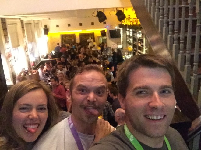
Monday night was also quite notable. During the trip I became quite involved with the EMS Wolfpack (@EMSWolfpack) and EMS Pocus (@emspocus, http://www.emspocus.com). The two groups collaborated to throw a pub night. We rented out an entire bar, The Trinity Inn, for a night of informal point-of-care ultrasound education, socialization, and alcohol consumption. We had no idea what to expect, nor did we know what was coming.

It started off with a few people learning the basics of POCUS (echo, RUSH, vascular access). But, before we knew it, the bar was full. It was impossible to even walk through it. In addition, Jim DuCanto (@jducanto) was there with his SALAD vomiting airway mannequin. This is probably the only documented instance of SALAD being performed in a bar; we are very proud of this. People were learning difficult airway techniques all throughout the night until the bar kicked us out.


However, the learning that was done was not the most remarkable point of the night. We had providers of all levels, from every continent, and from all specialities of critical care attend. People  networked and collaborated with each other in a format that I believe is unique to SMACC. As was spoken about in SMACCforce, there was a true flattening of the heirarchy where paramedics were engaging physicians, and vice versa, in relevant and important conversation. The night was an overwhelming success and next year we will need a bigger pub.

Tuesday was the first day of the main conference. There were so many incredible talks that it would take me days to summarize them so I will focus on the ones that impacted me the most. The conference started with an incredible opening with a light show and the new smaccDUB soundtrack. This was followed by the John Hinds plenary, a series of speakers with topics designed to commemorate John.


Perhaps the most inspirational was Scott Weingart’s (@emcrit) talk where he encouraged us all the meditate. Scott has recently focused a lot on the psychological aspects of critical care and this is one way he believes that we can all improve our mental health and resuscitation abilities. There was also a video commemorating John’s life and a talk from his partner. I think there were tears throughout the auditorium.

“I will not leave you.”

-John Hinds to all of his patients

“Don’t be a resus wanker.”

-Also John

The rest of day one consisted of concurrent sessions. First I attended the Oxygen and Stress session where we learned from geniuses, including John Myburgh (@JAMyburgh) and self-proclaimed sex god Mervyn Singer that supplemental oxygen is probably a bad thing in most cases and patients do well without it.


The second half of the day was the How I Do It session. Providers spoke on specific practices they use regularly. Casey Parker (@broomedocs) discussed the importance of using evidence based values such as pretest probability when performing tests. In general many of the tests done in the emergency department are not helpful and difficult to interpret. Per Bredmose spoke on his use of inhaled nitric oxide in Norwegian helicopter critical care. Their use focuses primarily on bridging ARDS patients to ECMO but also in any cause of pulmonary hypertension such as pulmonary embolism. The day concluded with a panel discussing how medical knowledge and new data should be disseminated. There was argument about peer-review, journal bias, and the high cost to access new studies. The broad consensus was that we need a centralized, open access location to put all new data which has a publicly viewable peer-review process.

Day two started with a plenary called Slaying Sacred Cows. This was a series of four incredible talks which truly all deserve individual recognition. Resa Lewis (@ultrasoundREL) spoke inspirationally about women as leaders. She taught lessons that are designed to help both men and women succeed as leaders in medicine.

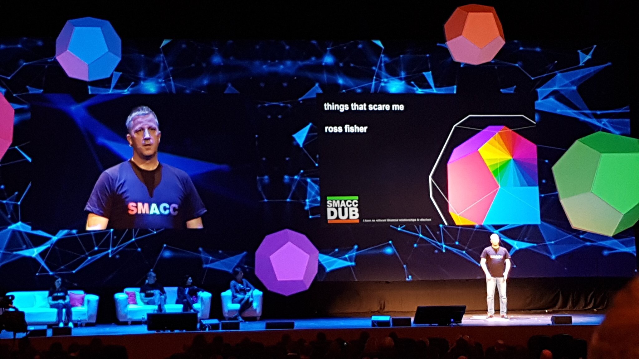
The talk that may have been the most impactful and lacrimation generating of the entire week was Ross Fisher’s (@ffolliet) Things That Scare Me, which he gave with no slides as the audience sat in intent silence.


Ross is a pediatric surgeon in the UK and he described some of the times during his practice that he has been most scared. This included operating on a 600 gram neonate with NEC. A patient this size has a blood volume of about 60 mL and they tend to bleed. He said that fear is the emotion that ends careers of excellent providers. The most important factor in dealing with fear is support from colleagues. In the right environment, fear can be overcome. This talk took such courage to give to a room of 2,000+. It is a must hear for everyone once it is released.

The next was from Reuben Strayer (@emupdates) who spoke about treatment of agitation. The most important part of the talk was this: we want droperidol back in the U.S.! It is a safe and effective drug for the significantly disruptive patient. If the patient is combative, or a threat to themselves or others, IM ketamine is the agent of choice.

The last talk of the morning plenary was from the amazing Liz Crowe (@LizCrowe2). She spoke about love in critical care and how it really impacts patient outcomes and provider health. As expected the talk was full of laughs and sexual enuendo. Remember your patients, patient families, and colleagues all need love.

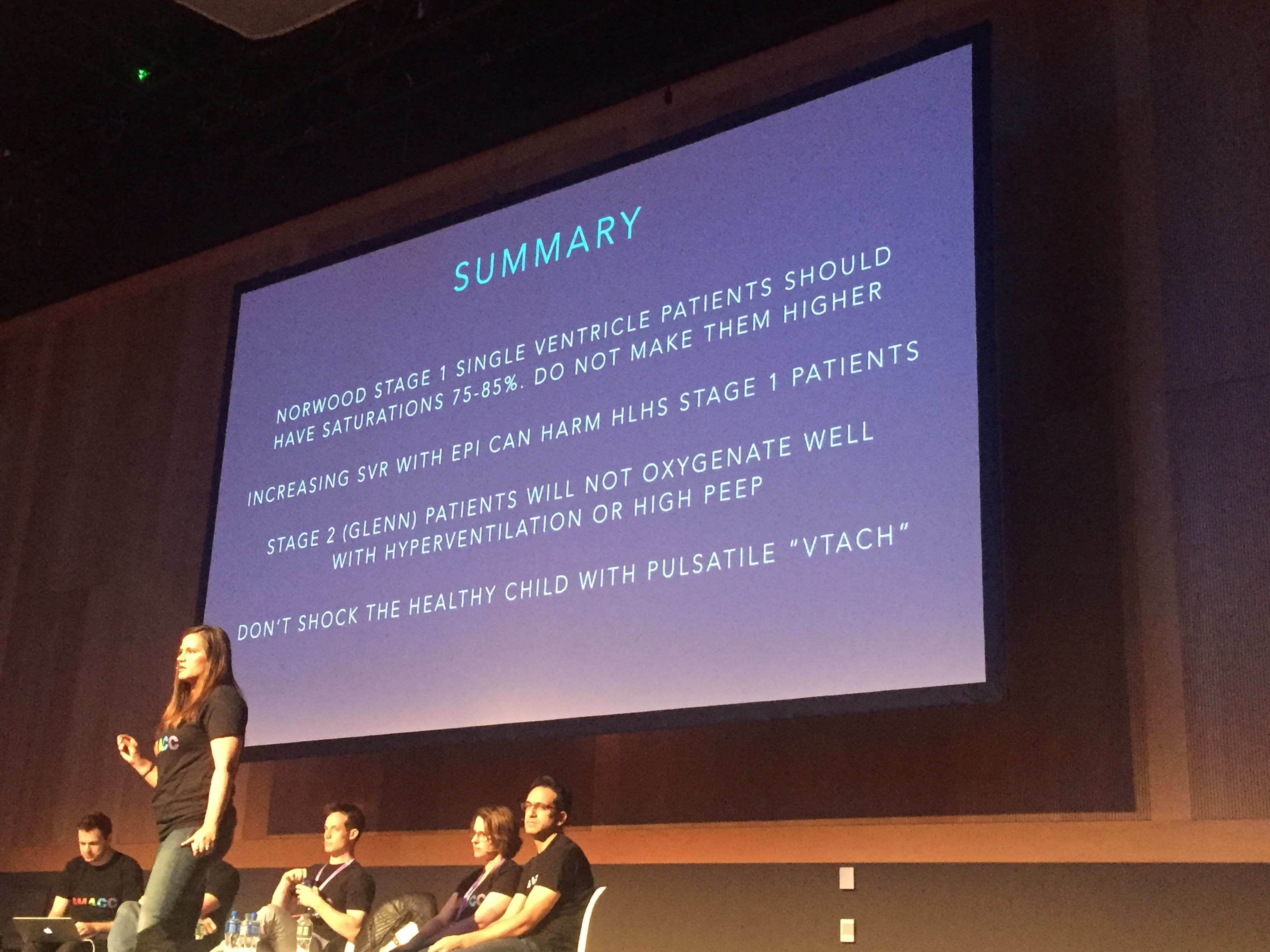
The morning concurrent for me was All Heart. There were awesome talks on pediatric heart conditions, cardiac surgery patients, and the RV.


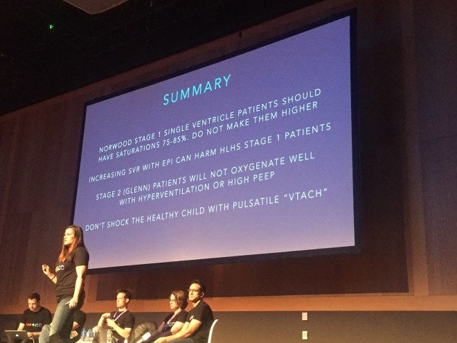
Haney Mallemat (@CriticalCareNow) intrigued the audience by proposing a modern approach to PEA arrest. Not all are the same and underlying causes must be identified and treated if ROSC is to occur. PEA is really an umbrella term, much like SVT. Ultrasound should be used to distiguish between pulselessness with rhythm and echo motion (PREM) and pulselessness with rhythm and echo standstill (PRES).

The afternoon of day two featured three different forums, of which I attended Bare Knuckle EM. There were four sessions which included an EM literature update. It turns out bacon can be used to treat epistaxis! Who knew? (http://www.ncbi.nlm.nih.gov/pubmed/22224315) Actual relevant topics were also discussed. The three following sessions were pro/con debates about important topics in EM. The topics were: EM is a failed paradigm, POCUS is a problem, and thrombolysis in submassive PE. Check these when they are released. The day concluded with SONIC BOOM, an entertaining and educational display of POCUS techniques.

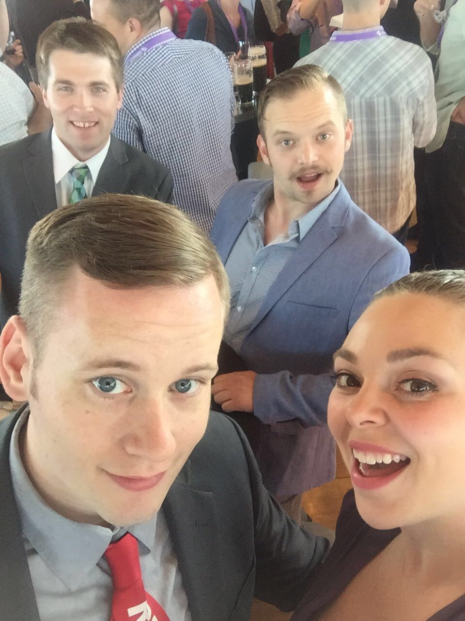
We can’t get through day two without mentioning the incredible gala. The event was held at the Guinness Storehouse, the five floor venue where Guinness is made and sold in large quantities. Around 2,100 people attended and it was an awesome time.


The last day started off interestingly. The auditorium was about 1/4 full as a result of gala activities the night before. It gradually filled throughout the morning. The plenary talks were themed Beyond the Ivory Towers. The first two talks were from Kath Maitland (@KathMaitland) and Nikki Blackwell. They both discussed topics regarding their work in Africa providing care for critically ill patients with few resources. Kath focused on oxygen and its potential overuse.

Next, EM physcian Ashley Shreves discussed the immensely important topic of palliative care in the ED. This is done poorly pretty much everywhere. Incredible talk that will inspire you to do better.

“The treatment for death is probably not CPR in many cases!”

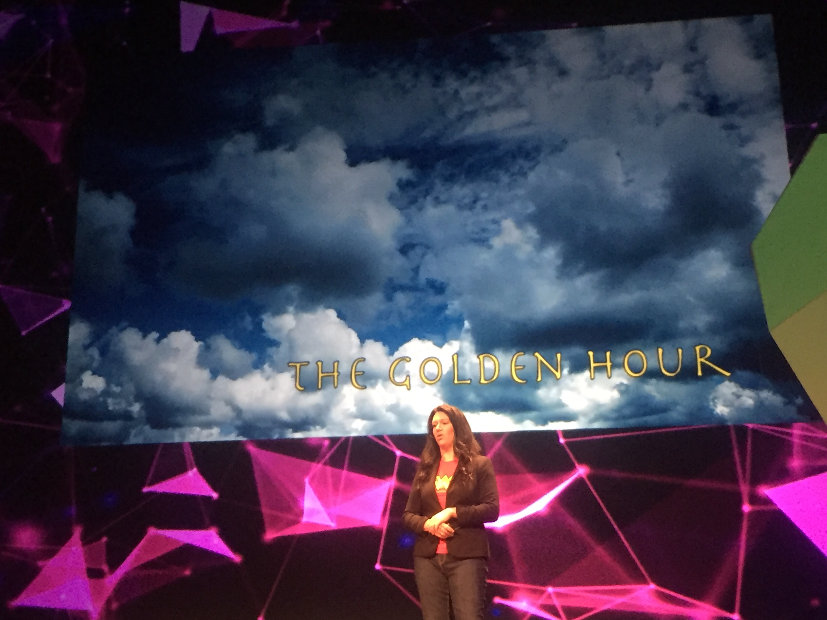
Ashley Liebig (@ashleyliebig) stole the show with her talk The Golden Fleece, The Golden Hour, and The Golden Rule.


In critical care, pre-hospital and in-hospital, providers carry a massive burden. Clinical responsibility paired with emotional trauma often leads to the downfall of incredible people. Ashley gave an emotional and honest talk with essentially no slides. I encourage everyone to listen to this when it is released.

Brining Sexy Back was the theme of my morning session. Weingart spoke about post-intubation sedation and analgesia. Remember: analgesia always and FIRST. The most notable talk though was from Alex Psirides (@psirides) on Why Your Hospital is Broken.


This talk was a reality check but true for most places. The focus of most hospital systems is often not truly the patient, it is the providers. Another must-hear.

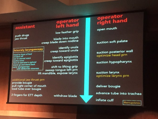
My last concurrent of the conference was A is for Airway. Four great talks here. Reuben Strayer presented a somewhat novel approach to laryngoscopy designed to make providers slow down and not rush. Listen to the lecture and see what its all about.

The finale was a ton of fun. Two teams of four were pitted against eachother in contests testing knowledge, teamwork, and skills. All the participants earned half price admission to SMACC next year!

So in summary: epic. This post does not even scratch the surface. The talks were incredible. But what really makes SMACC special is the people you meet. And I can’t quanitify that in words. If you have not been to SMACC, go! Also, if you are not involved in #FOAMed and the twitter world I encourage you to start. Go to http://www.smacc.net.au and listen to the previous talks. Also, go to http://www.lifeinthefastlane.com, the original FOAMed site which has thousands of articles. Get on twitter, search #FOAMed, #smaccDUB, and get involved.
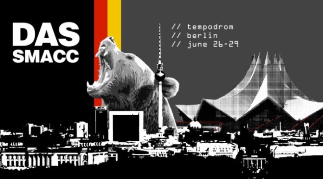

#dasSMACC 2017

Next year SMACC will be held in Berlin, Germany.

Venue: Tempodrom

June 26-29, 2017

#smaccDUB reflection